
Real-time tumor tracking radiotherapy
J Appl Clin Med Phys. 2016 Jul 8;17(4):202-213
Phys Med Biol. 2017 Feb 21;62(4):1585-1599
Phys Med Biol. 2018 Mar 21;63(6):065016.
Lung and liver tumors with respiratory movement have been treated by including the area of the tumor that moves with respiration in the irradiation range, but this has resulted in high radiation doses to normal organs and increased side effects. To solve this problem, we have introduced a dynamic tracking radiotherapy system to provide high-precision radiotherapy. We are developing methods and software for the safe introduction of these technologies into clinical practice.
Tumor Tracking Algorithm
Markerless tumor tracking
Most of the current respiratory migration therapies use X-ray images to recognize the location of tumors and to treat them. However, implanting a metal marker in the body is burdensome to patients. Therefore, we are developing an algorithm to recognize the tumor directly using image processing.
Templateless marker tracking (A joint study by Shimadzu Corporation)
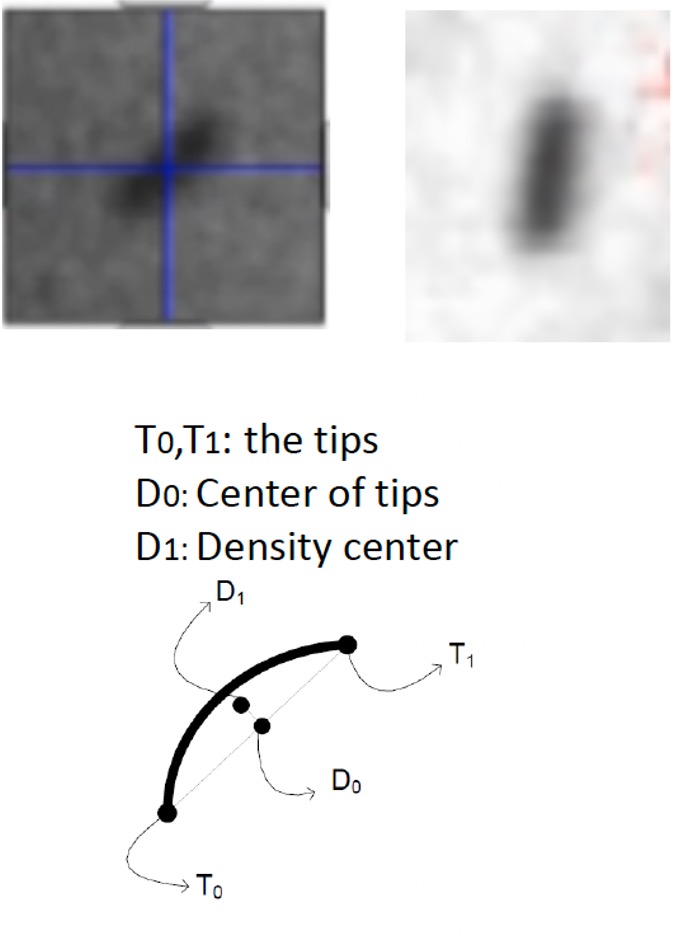
There are various shapes of metal markers that are implanted near the tumor. Markers implanted in the liver are coiled, and the coil shape changes depending on the direction of fluoroscopy. Therefore, conventional marker tracking methods require the preparation of a template for each direction of fluoroscopy, which increases treatment time. We have developed an algorithm to track coil-shaped markers without using templates.
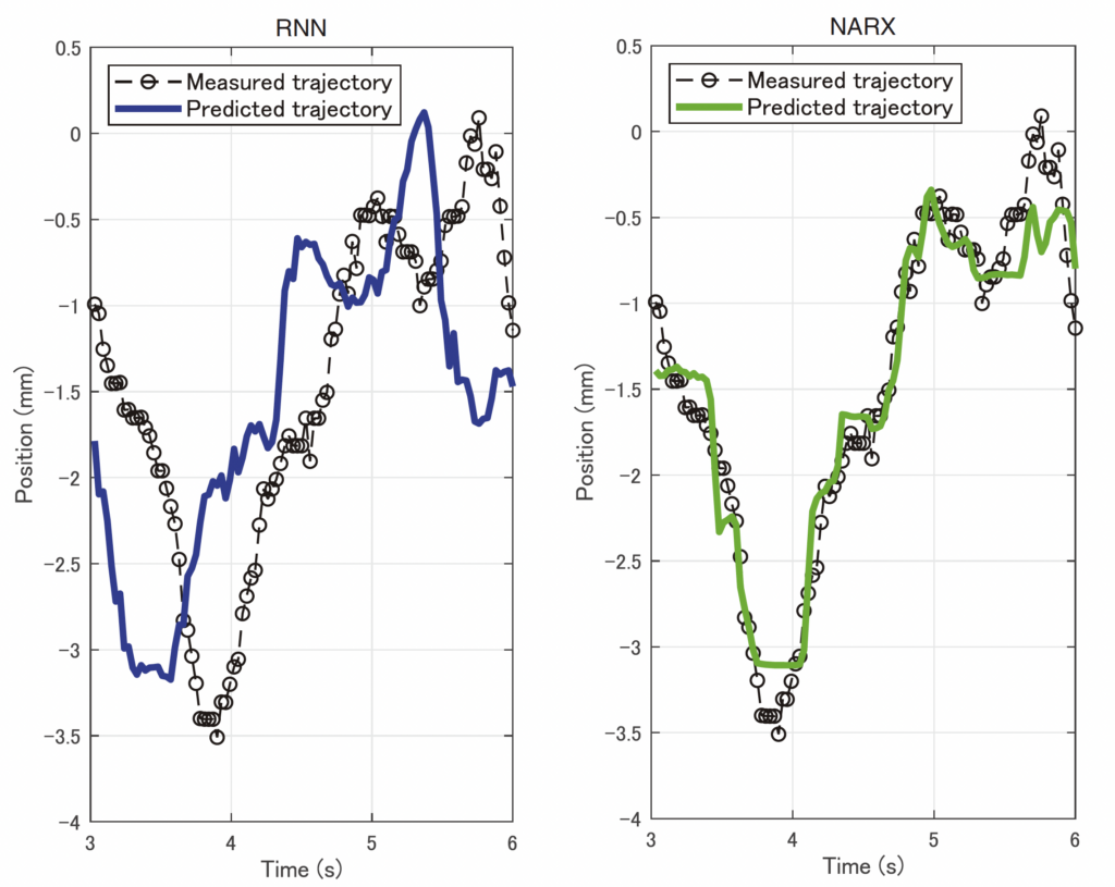
Tumor location prediction model
Phys Med Biol. 2019 Oct 31;64(21):21NT02.
Real-time tumor-tracking therapy or dynamic tracking irradiation involves a mechanical delay because the treatment is performed after the tumor is recognized. This delay has a significant impact on treatment accuracy. To compensate for this delay, we are developing an algorithm that compensates for delay by predicting the tumor position.
Reduction of the fluoroscopy dose
View 1
View 2
Real-time tumor-tracking radiotherapy uses fluoroscopic images to recognize the movement and position of the tumor. The use of fluoroscopic images during treatment for a long period of time increases the radiation dose. We have developed an algorithm to reconstruct a pseudo-high-dose fluoroscopic image from a low-dose fluoroscopic image (Shiinoki et al. (Shiinoki et al. AAPM 2019)
Works
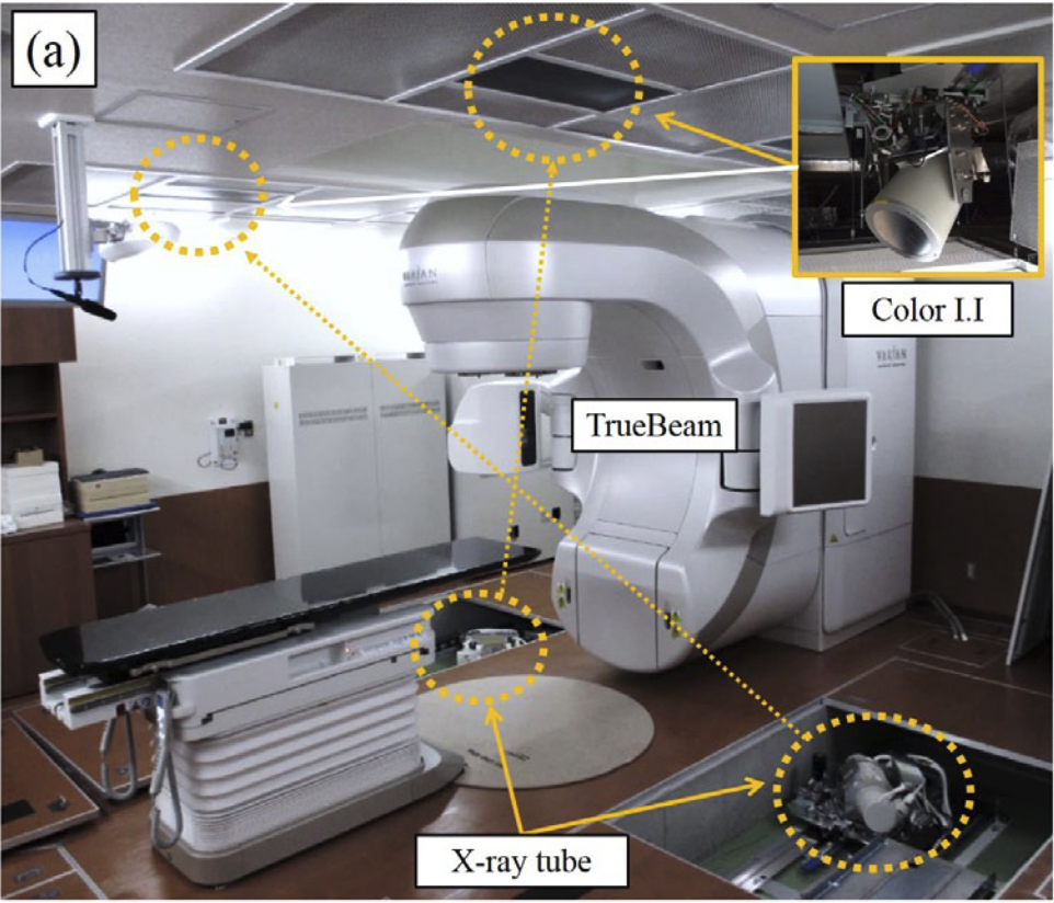
- Evaluation of a combined respiratory-gating system comprising the TrueBeam linear accelerator and a new real-time tumor-tracking radiotherapy system: a preliminary study.
Shiinoki T, Kawamura S, Uehara T, Yuasa Y, Fujimoto K, Koike M, Sera T, Emoto Y, Hanazawa H, Shibuya K.
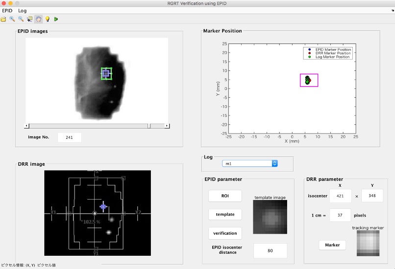
J Appl Clin Med Phys. 2016 Jul 8;17(4):202-213. Erratum in: J Appl Clin Med Phys. 2017 Jul;18(4):238. - Verification of respiratory-gated radiotherapy with new real-time tumour-tracking radiotherapy system using cine EPID images and a log file.
Shiinoki T, Hanazawa H, Yuasa Y, Fujimoto K, Uehara T, Shibuya K.
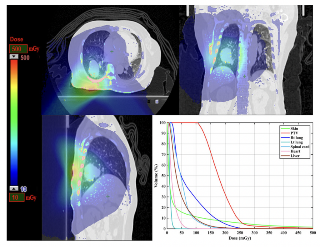
Phys Med Biol. 2017 Feb 21;62(4):1585-1599. - Estimation of patient-specific imaging dose for real-time tumour monitoring in lung patients during respiratory-gated radiotherapy.
Shiinoki T, Onizuka R, Kawahara D, Suzuki T, Yuasa Y, Fujimoto K, Uehara T, Hanazawa H, Shibuya K.
Phys Med Biol. 2018 Mar 21;63(6):065016. - Evaluation of the effects of motion mitigation strategies on respiration-induced motion in each pancreatic region using cine-magnetic resonance imaging.
Fujimoto K, Shiinoki T, Yuasa Y, Onizuka R, Yamane M.
J Appl Clin Med Phys. 2019 Sep;20(9):42-50. - Prediction of lung tumor motion using nonlinear autoregressive model with exogenous input.
Jiang K, Fujii F, Shiinoki T.
Phys Med Biol. 2019 Oct 1. doi: 10.1088/1361-6560/ab49ea.
Research Grants
- 超高線量率四次元動体追跡照射の高精度化に向けた新しい品質管理法の開発
文部科学省: 科学研究費補助金(若手研究(B)) - 四次元動体追跡治療のための新型動体追跡装置による透視被ばく線量計算システムの開発
がん研究振興財団: がん研究助成(A) - 四次元動体追跡放射線治療のための金属マーカを使用しない非侵襲的肺腫瘍追跡技術の開発
新日本先進医療研究財団: がん研究助成 - 深層学習を用いた肺腫瘍判別によるマーカーレス四次元動体追跡放射線治療の確立
文部科学省: 科学研究費補助金(基盤研究(C)) - ビジュアルバイオフィードバックシステムを用いた動体追跡強度変調放射線治療の確立
文部科学省: 科学研究費補助金(基盤研究(C)) - バイオメカニクスとロボティクスに基づく肺機能/形態融合の次世代動体追跡放射線治療
文部科学省: 科学研究費補助金(基盤研究(C)) - 膵癌の呼吸性移動特性の定量化のための深層学習を用いた新しい四次元MRI構築法の開発
文部科学省: 科学研究費補助金(奨励研究)